Single-Cell Neuroscience

For many years, understanding the brain's complex workings has been a challenge for scientists. The brain consists of numerous cell types with intricate communication networks. Traditional methods analyze brain tissue as a whole, missing the unique contributions of individual cells. This is similar to analyzing an orchestra's sound without distinguishing the instruments or musicians.

Single-cell neuroscience is a groundbreaking field that allows researchers to examine the brain at the level of single neurons. By isolating and analyzing the genetic makeup (transcriptome) of individual cells, scientists can gain a deeper understanding of cellular diversity, function, and their roles in both healthy and diseased brains.

Pioneering Techniques:

A key technology driving single-cell neuroscience is single-cell RNA sequencing (scRNA-seq). This technique allows researchers to measure messenger RNA (mRNA) molecules within a single cell. mRNA acts as a blueprint for protein synthesis. By identifying the mRNA profile, scientists can determine the genes actively expressed in that particular cell. This information provides valuable insights into the cell's identity and function.

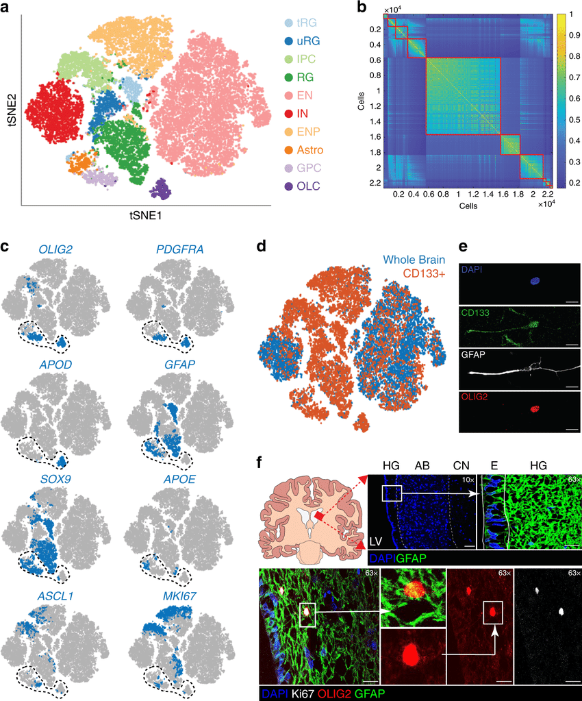
Single-cell RNA sequencing of the developing brain and the identification of glial progenitor cells a T-distributed stochastic neighbour embedding (tSNE) map of human fetal brain cells by cluster or cell type. Data sets from total cells and CD133+ cells were combined. Cells are colored by cell type. tRG truncated radial glia, uRG unknown radial glia, IPC inhibitory neuronal progenitor, RG radial glia, EN excitatory neuron, IN interneuron, ENP excitatory neuronal progenitor, Astro astrocyte, GPC glial progenitor cell, OLC oligo-lineage cells. b Similarity matrix of fetal brain cells ordered by cluster. c tSNE maps of human fetal brain cells showing cell type expression of OLIG2, PDGFRA, APOD, GFAP, SOX9, APOE, ASCL1, and MKI67. Expression is averaged to the 20 closest neighbors in principal component (PC) space. Encircled cells were reclustered to yield three separate clusters. d tSNE map of total human fetal brain cells and CD133+ fetal brain cells. e Representative example of freshly cultured fetal neural stem cells coexpressing CD133, OLIG2, and GFAP (n = 2 independent biological samples). Images were taken at ×63 magnification. Scale bars: 10 μmf Immunofluorescence analysis of the adult human subventricular zone (SVZ) at the junction of the AB and HG. Top row, schematic and anatomic structure of the SVZ. Bottom row, identification of dividing cells with marker expression corresponding to glial progenitor cells. HG hypocellular gap, AB astrocytic band, E ependymal cells, LV lateral ventricle, CN caudate nucleus. Analysis was performed in n = 4 independent patient samples. Scale bars: top row images: 200 μm (left) and 40 μm (right); bottom row images: 20 μm.

Unveiling Cellular Diversity:

The brain is composed of specialized cell types, each with a specific role in neural circuits. Single-cell analysis has revealed a surprising level of variation within these populations. For instance, studies have identified distinct subtypes of neurons within the cortex, each with unique gene expression patterns that likely contribute to specific functions such as sensory processing or memory formation.

Selected relevant scRNA-seq studies revealing brain heterogeneity. Recent high throughput brain scRNA-seq studies indicate that mouse brain is composed of a large diversity of specialized cell subpopulations. Arrows indicate the sample collection region and the number of isolated cells. The numbers to the left represent the quantity of cells belonging to each global cell type. The numbers to the right represent the quantity of subpopulations found within each global cell type. Asterisks indicate cells were enriched for oligodendrocyte-lineage.

Decoding Brain Function:

By linking specific gene expression profiles to neuronal function, researchers are gaining a clearer understanding of how different cell types contribute to brain circuits and behavior. Companies like Gentaur Group are playing a very important role in this progress by providing high-quality reagents essential for scRNA-seq experiments. For instance, scRNA-seq has been used to identify specific neuronal populations involved in learning and memory. This knowledge is crucial for developing targeted therapies for neurological disorders that affect these processes.


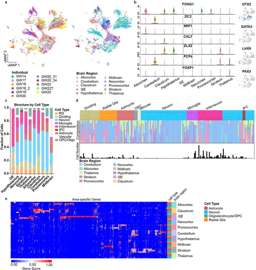
Single-cell analysis across the whole developing human brain a, UMAP plots showing the representation of samples by individual and brain region. There is strong intermixing across individuals, but more segregation by stage. b, On the left, violin plots of known and novel brain region-enriched marker genes. On the right are feature plots of cell type specific transcription factors. c, Histogram depicting the cell type composition as determined by the single-cell analysis for each sampled brain region, showing similar distributions across regions, but with known enrichments for inhibitory interneurons in the ganglionic eminences, and other small enrichments for specific cell types in other regions. d, Hierarchical clustering of 210 neocortex clusters based upon Pearson correlations across cluster markers. Each bar is colored based upon the major cell type assigned to that cluster. Beneath the clusters are histograms showing the fraction of cells from each area contributing to the cluster, and below that is a barchart showing the relative number of cells in each cluster (log2 transformed numbers ranging from 0 to 20). e, Heatmap representing the universe of area-specific genes for each cell type. Gene score, a metric that combines specificity and fold change, is shown from blue to red. Rows are grouped by brain region, and reveal that in many structures, area-specific genes cross cell types.

Illuminating Disease Mechanisms:

Single-cell approaches are transforming our understanding of neurological diseases. By comparing the transcriptomes of healthy and diseased cells, researchers can pinpoint the specific cell types most affected by the pathology. This knowledge can help identify new therapeutic targets and pave the way for the development of personalized medicine strategies for neurological disorders like Alzheimer's disease and Parkinson's disease.

Conclusion:

Single-cell neuroscience is revolutionizing our understanding of the brain. By studying the brain one cell at a time, researchers are unraveling the mysteries of cellular diversity, function, and disease. This knowledge holds immense promise for the development of novel therapies and personalized medicine approaches for neurological disorders, ultimately paving the way for a brighter future for brain health.

For additional information, you may watch this video:

in News
Single-Cell Neuroscience
Gen store June 4, 2024
Tags
Sign in to leave a comment
Neurotransmitters: Orchestrating Communication Between Neurons