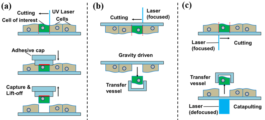
The brain is a complex organ composed of many different cell types, each with a specific function. These cells work together to allow us to think, move, and experience the world around us. Traditionally, scientists have studied the brain by analyzing tissue samples containing many cells at once. This approach provides limited information because it masks the unique proteins present in each cell type.
Single-cell proteomics is a new technique that allows scientists to directly analyze the proteins within individual brain cells. This provides a much more detailed picture of how different brain cells function.
Challenges and Techniques
One of the main challenges in single-cell proteomics is that there are very few proteins in each cell. Scientists have overcome this hurdle by developing techniques to isolate individual cells from brain tissue. These techniques include laser capture microdissection (LCM) and fluorescence-activated cell sorting (FACS).
Once isolated, the proteins in the cell are broken down into smaller molecules called peptides. These peptides are then analyzed by a machine called a mass spectrometer. The mass spectrometer acts like a fingerprint reader, identifying and counting the different peptides present. By analyzing the peptides, scientists can determine the complete set of proteins, or proteome, of each cell.
High-quality reagents optimized for FACS are crucial for this process. Companies like Maxanim offer a variety of solutions for researchers in this field.
Benefits and Future Applications
Single-cell proteomics has the potential to revolutionize our understanding of the brain. It allows scientists to:
- Identify different cell types: By analyzing the protein profiles of individual cells, scientists can create more accurate classifications of brain cell types.
- Map protein location: New techniques can visualize where proteins are located within a cell, providing insights into cellular organization and function.
- Understand disease: By comparing the proteomes of healthy and diseased brain cells, scientists can identify proteins that are involved in neurodegenerative disorders such as Alzheimer's and Parkinson's disease.
The field of single-cell proteomics is constantly evolving. As technology improves, scientists will be able to detect a wider range of proteins from individual cells and map their location with even greater precision. By combining single-cell proteomics with other techniques, scientists will gain a deeper understanding of how brain cells function in health and disease, paving the way for the development of new therapies for neurological disorders.
Learn more about Spatial and Single Cell Genomics in this video:

Single-Cell Proteomics in Brain Research